Author Affiliations
Abstract
1 Department of Orthopaedics, The First Affiliated Hospital of Shantou University Medical College Shantou 515041, Guangdong Province, P. R. China
2 MOE Key Laboratory of Laser Life Science & Institute of Laser Life Science College of Biophotonics, South China Normal University Guangzhou 510631, P. R. China
3 Guangdong Provincial Key Laboratory of Laser Life Science College of Biophotonics, South China Normal University Guangzhou 510631, P. R. China
4 Guangzhou Key Lab of Spectral Analysis and Functional Probes College of Biophotonics, South China Normal University Guangzhou 510631, P. R. China
Bioprobe based on fluorescence is widely used in biological and medical research due to its high sensitivity and selectivity. Yet, its quantification in vivo is complicated and often compromised by the interaction between the fluorophore with the environmental factors, as well as the optical scattering and absorption by the tissue. A high florescence quantum yield and minimal interference by the environment are key requirements for designing an effective bioprobe, and the pre-requisitions severely limit the available options. We propose that a comprehensive evaluation of potential bioprobe can be achieved by simultaneously measuring both radiative and non-radiative transitions, the two fundamental and complementary pathways for the energy de-excitation. This approach will not only improve the accuracy of the quantification by catching the information from a broader spectrum of the energy, but also provide additional information of the probe environment that often impacts the balance between the two forms of the energy transition. This work first analyzes the underlying mechanism of the hypothesis. The practical feasibility is then tested by means of simultaneous measurements of photoacoustic signal for the non-radiative and fluorescence for the radiative energy processes, respectively. It is demonstrated that the systematic evaluation of the probe energy de-excitation results in an improved quantitative tracing of a bioprobe in complex environment.
Bioprobe fluorescence photoacoustic Journal of Innovative Optical Health Sciences
2023, 16(4): 2243002
Author Affiliations
Abstract
1 MOE Key Laboratory of Laser Life Science & Institute of Laser Life Science, College of Biophotonics, South China Normal University, Guangzhou 510631, P. R. China
2 Guangdong Provincial Key Laboratory of Laser Life Science, College of Biophotonics, South China Normal University, Guangzhou 510631, P. R. China
3 Guangzhou Key Lab of Spectral Analysis and Functional Probes, College of Biophotonics, South China Normal University, Guangzhou 510631, P. R. China
As an emerging hybrid imaging modality, microwave-induced thermoacoustic imaging (MTAI), using microwaves as the excitation source and ultrasonic signals as the information carrier for combining the characteristics of high contrast of electromagnetic imaging and high resolution of ultrasound imaging, has shown broad prospects in biomedical and clinical applications. The imaging contrast depends on the microwave-absorption coefficient of the endogenous imaged tissue and the injected MTAI contrast agents. With systemically introduced functional nanoparticles, MTAI contrast and sensitivity can be further improved, and enables visualization of biological processes in vivo. In recent years, functional nanoparticles for MTAI have been developed to improve the performance and application range of MTAI in biomedical applications. This paper reviews the recent progress of functional nanoparticles for MTAI and their biomedical applications. The challenges and future directions of microwave thermoacoustic imaging with functional nanoparticles in the field of translational medicine are discussed.As an emerging hybrid imaging modality, microwave-induced thermoacoustic imaging (MTAI), using microwaves as the excitation source and ultrasonic signals as the information carrier for combining the characteristics of high contrast of electromagnetic imaging and high resolution of ultrasound imaging, has shown broad prospects in biomedical and clinical applications. The imaging contrast depends on the microwave-absorption coefficient of the endogenous imaged tissue and the injected MTAI contrast agents. With systemically introduced functional nanoparticles, MTAI contrast and sensitivity can be further improved, and enables visualization of biological processes in vivo. In recent years, functional nanoparticles for MTAI have been developed to improve the performance and application range of MTAI in biomedical applications. This paper reviews the recent progress of functional nanoparticles for MTAI and their biomedical applications. The challenges and future directions of microwave thermoacoustic imaging with functional nanoparticles in the field of translational medicine are discussed.
Microwave thermoacoustic imaging nanomaterials nanoprobe Journal of Innovative Optical Health Sciences
2023, 16(2): 2230014
Author Affiliations
Abstract
1 MOE Key Laboratory of Laser Life Science & Institute of Laser Life Science, College of Biophotonics, South China Normal University, Guangzhou 510631, China
2 Guangdong Provincial Key Laboratory of Laser Life Science, College of Biophotonics, South China Normal University, Guangzhou 510631, China
3 Guangzhou Key Laboratory of Spectral Analysis and Functional Probes, College of Biophotonics, South China Normal University, Guangzhou 510631, China
4 e-mail: xingda@scnu.edu.cn
5 e-mail: qinghuan@scnu.edu.cn
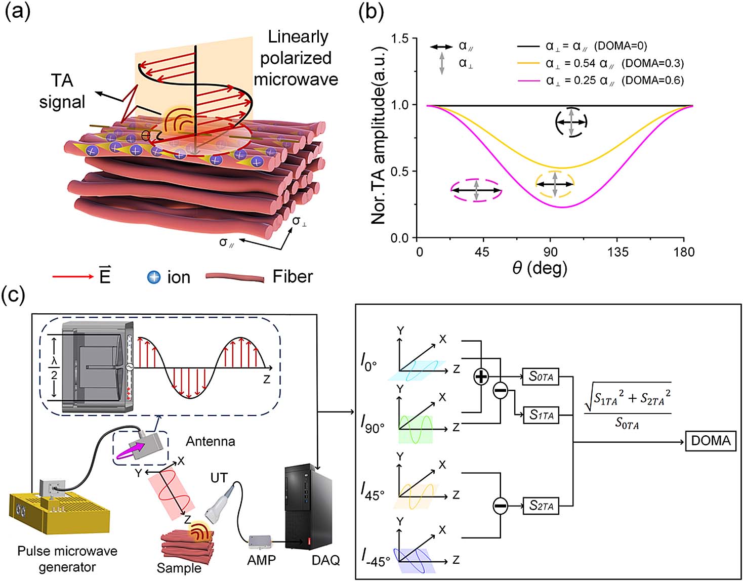
Polarization optical imaging can be used to characterize anisotropy in biological tissue microstructures and has been demonstrated to be a powerful tool for clinical diagnosis. However, the approach is limited by an inability to image targets deeper than due to strong optical scattering in biological tissues. As such, we propose a novel polarization microwave-induced thermoacoustic imaging (P-MTAI) technique to noninvasively detect variations in deep tissue by exploiting the thermoacoustic signals induced by four pulsed microwaves of varying polarization orientations. The proposed P-MTAI method overcomes the penetration limits of conventional polarization optical imaging and provides submillimeter resolution over depths of several centimeters. As part of the paper, the structural characteristics of tissues were quantified using a new parameter, the degree of microwave absorption anisotropy. P-MTAI was also applied to the noninvasive detection of morphological changes in cardiomyocytes as they transitioned from ordered to disordered states, providing a potential indication of myocardial infarction.
Photonics Research
2022, 10(5): 05001297
1 华南师范大学激光生命科学教育部重点实验室,广东 广州 510631
2 广东省激光生命科学重点实验室,广东 广州 510631
3 广州市光谱分析与功能探针重点实验室,广东 广州 510631
4 华南师范大学生物光子学研究院,广东 广州 510631
微波热声成像(MTAI)结合微波成像高对比度和超声成像高分辨率的优势,是一项新型的无损物理医学成像方法。MTAI利用微波作为激发源,超声作为信息载体,通过微波到超声能量传递形式的变换,实现生物组织无损、厘米深度的高分辨率成像。MTAI对比度取决于微波吸收差异,在生物组织中主要以水分子等极性分子(分子极化损耗)和离子(离子极化损耗)作为信号来源,从而获得生物组织的结构和功能图像,在生物医学成像领域具有独特的优势,得到了众多科研工作者的广泛关注。本综述从MTAI技术的原理、微波辐射组件、数据采集组件和数据处理组件、MTAI技术在生物医学领域的应用以及MTAI探针等方面进行描述,并结合当前MTAI技术面临的挑战对未来的发展方向进行了展望。
激光与光电子学进展
2022, 59(6): 0617004
红外与激光工程
2021, 50(11): 20210551
Author Affiliations
Abstract
Ministry of Education Key Laboratory of Laser Life Science and Institute of Laser Life Science, College of Biophotonics, South China Normal University, Guangzhou 510631, P. R. China
Photoacoustic imaging (PAI) breaks through the optical diffusion limit by making use of the PA effect. By converting incident photons into ultrasonic waves, PAI combines high contrast of optical imaging and high spatial resolution in depth tissue of ultrasound imaging in a single imaging modality. This imaging modality has now shown potential for molecular imaging, which enables visualization of biological processes with systemically introduced functional nanoparticles. In the current review, the potentials of different optical nanoprobes as PAI contrast agents were elucidated and discussed.
Photoacoustic imaging optical nanoprobes molecular imaging Journal of Innovative Optical Health Sciences
2017, 10(4): 1730004
华南师范大学激光生命科学研究所, 激光生命科学教育部重点实验室, 生物光子学研究院, 广东 广州 510631
光声成像技术是近年来发展的一种新型的无损医学成像技术, 它是以脉冲激光作为激发源, 以检测的声信号为信息载体, 通过相应的图像重建算法重建组织内部结构和功能信息的成像方法。该方法结合了光学成像和声学成像的特点, 可提供深层组织高分辨率和高对比度的组织层析图像, 在生物医学临床诊断以及在体成像领域具有广泛的应用前景。目前光声成像的扫描方式主要有基于步进电机扫描方式和基于振镜的扫描方式, 本文针对目前步进电机扫描速度慢 (10 mm×10 mm; 0.001 帧/s), 振镜扫描范围小(<1 mm2)的不足, 发展了基于直线电机扫描的大视场快速光声显微成像系统。同一条扫描线过程中直线电机速度最高可达200 mm/s。 该技术采用逐线采集光声信号的方式, 比逐点采集光声信号的步进电机快800倍。该系统对10 mm×10 mm 全场扫描的扫描速度为0.8 帧/s。最大可扫描视场范围可以达到50 mm×50 mm。大视场快速光声显微成像系统的发展将为生物医学提供新的成像工具。
光声成像 直线电机 光声显微成像 photoacoustic imaging linear motor photoacoustic microimaging
华南师范大学激光生命科学研究所 激光生命科学教育部重点实验室, 生物光子学研究院, 广东 广州 510631
无损光声成像技术结合了纯光学成像高选择特性和纯超声成像中深穿透特性的优点, 克服了光散射限制, 实现了对活体深层组织的高分辨、高对比度成像。该成像技术对内源物质例如脱氧血红蛋白、含氧血红蛋白、黑色素、脂质等进行成像, 提供了活体生物组织结构和功能信息, 已经在生物医学领域表现出巨大的应用前景。然而, 很多与病理过程相关的特征分子的光吸收能力较弱, 在活体环境中难以被光声成像系统所识别, 从而限制了光声成像技术的应用范围。基于功能纳米探针的光声成像-光声分子成像极大拓展光声成像的应用范围, 可以在活体层面对病理过程进行分子水平的定性和定量研究, 将为实现目标疾病的早期诊断提供强大的技术支持。本文发展在近红外具有窄吸收线宽(半高宽仅为60 nm)的纳米金锥作为新型的光声探针。通过选择不同径长比的纳米金锥, 可以任意调节纳米金锥的吸收峰。通过调谐激光器的波长, 可实现对不同吸收峰纳米金锥的选择性激发。纳米金锥将有可能用于多光谱光声成像, 实现对不同靶标的目标分子探测。
无损光声成像 纳米金锥 多光谱光声成像 non-invasive photoacoustic imaging gold nanobipyramids multispectral photoacoustic imaging
华南师范大学生物光子学研究院激光生命科学研究所、暨激光生命科学教育部重点实验室, 广东 广州 510631
报道了一种利用单一波长激发的同时产生光声和荧光信号的显微成像系统, 本成像系统具有超高的成像分辨率(<6 μm)。借助外源的造影剂在近红外的吸收特性, 利用光声-荧光显微成像系统对活体肿瘤进行光声/荧光成像。实验结果表明, 光声-荧光显微镜在早期肿瘤的成像和检测等方面具有潜在的应用价值。因此, 通过研究和选择适当的双模态造影剂, 该系统在不同病理模型中可以提供更准确的组织信息及生理参数。
光声-荧光显微镜 活体肿瘤成像 造影剂 photoacoustic-fluorescence microscopy in vivo tumor imaging contrast agent
华南师范大学激光生命科学研究所 激光生命科学教育部重点实验室, 生物光子学研究院, 广东 广州 510631
多尺度显微成像系统(M-PAM)被发展, 并被用于成像从癌细胞到实体肿瘤的多尺度生物结构。该装置由二维运动平台,扫描振镜,物镜,聚焦超声换能器组成, 其横向分辨率达到3 μm。结果显示该系统可以对体外培养黑色素瘤细胞与体内的黑色素瘤进行无标记成像。基于具有靶向性的探针, M-PAM系统可以对体外培养的U87-MG肿瘤细胞以及体内U87-MG实体肿瘤进行成像。综上所述, M-PAM系统将是研究肿瘤的有力工具。
光声成像 肿瘤成像 多尺度成像 photoacoustic imaging tumor imaging multiscale imagin


